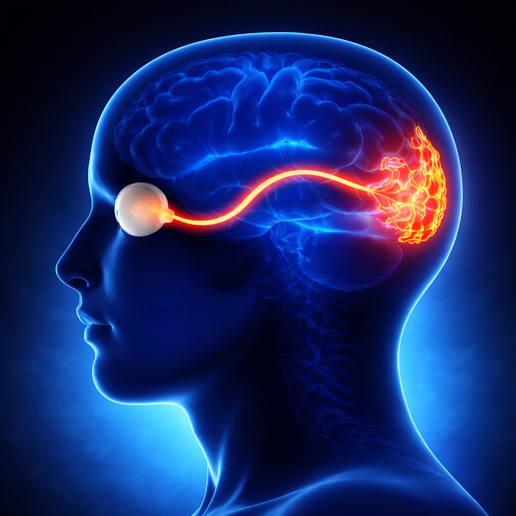
Optic neuropathies refer to a group of conditions affecting the optic nerve, which is responsible for transmitting visual information from the retina to the brain.
Continue reading to better understand the causes, symptoms, and treatments of the different types of optic neuropathies.
What does “optic neuropathy” mean?
Optic neuropathy is a condition affecting the optic nerve, with severity depending on the extent of nerve damage. Vision loss may occur gradually or suddenly, depending on the underlying mechanism.
Optic neuropathies can lead to:
reduced visual acuity
loss of central or peripheral vision
and in severe cases, sometimes even blindness.
Optic neuropathies may be bilateral (affecting both eyes) or unilateral (affecting one eye only).
The causes of optic neuropathies are varied.
The optic nerve plays a key role in visual function: any damage to its fibres can disrupt the transmission of visual signals to the cerebral cortex.
What are the inflammatory, vascular, and hereditary causes of optic neuropathies?
Optic neuropathies can be caused by inflammatory conditions such as optic neuritis, vascular disorders such as ischemia or hypertension, and hereditary factors.
Some optic neuropathies are associated with systemic diseases, such as multiple sclerosis, in which the nervous system is attacked, leading to degeneration of nerve fibres and the optic nerve.
In advanced stages of optic neuropathy, optic atrophy may develop, reflecting irreversible damage to nerve fibres.
Disease progression depends closely on the underlying cause and how quickly treatment is initiated.
For example, ischemic optic neuropathy results from a sudden reduction in blood supply to the optic nerve.
It occurs more frequently in individuals with cardiovascular risk factors (diabetes, hypertension, lipid disorders).
Hereditary forms may appear earlier and often require family screening.
What ophthalmic tests and imaging are used for optic neuropathies?
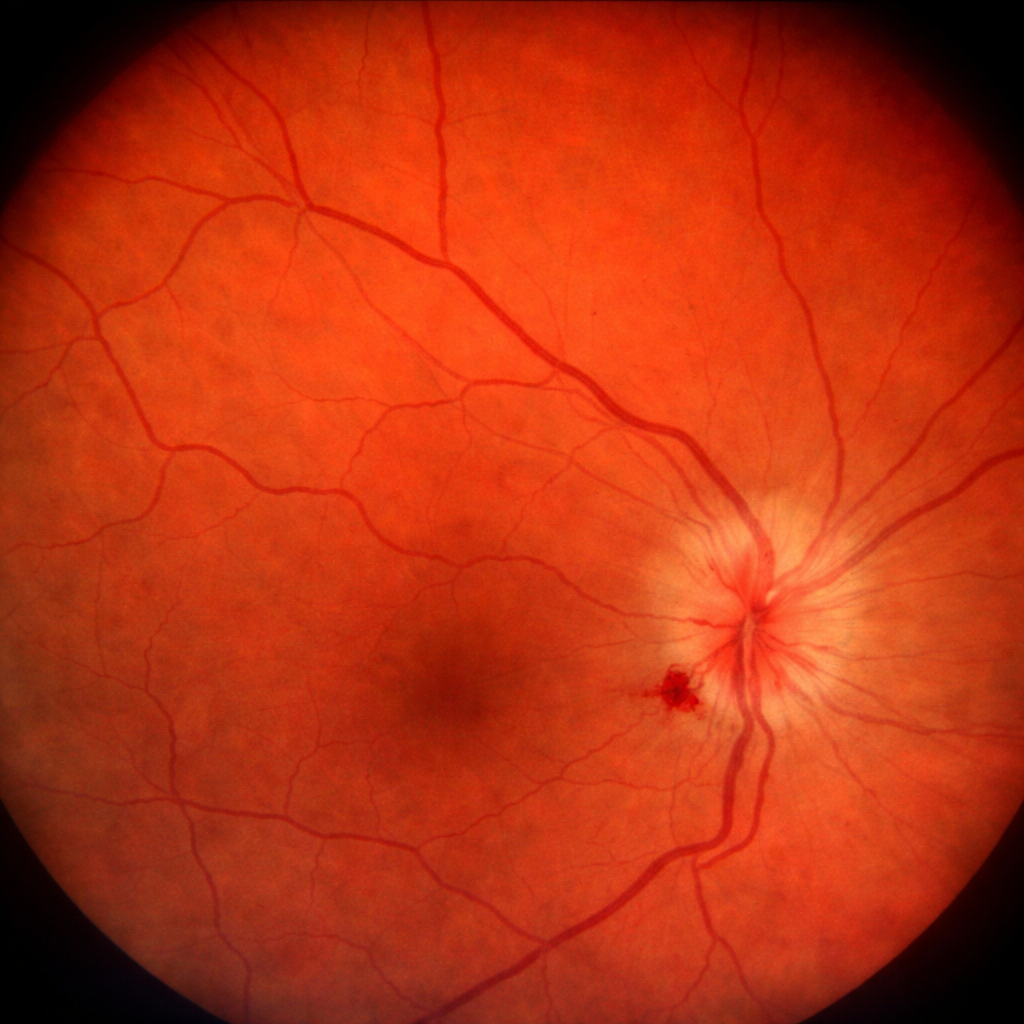
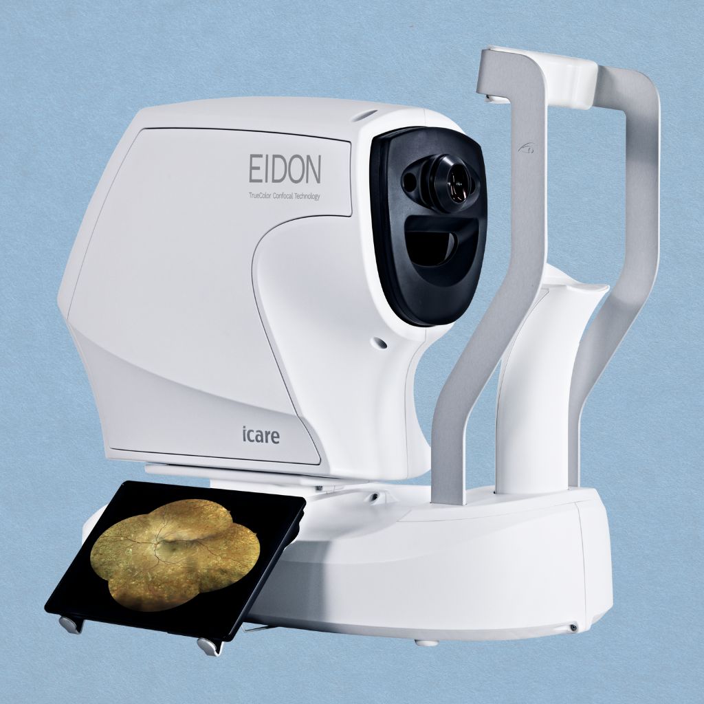
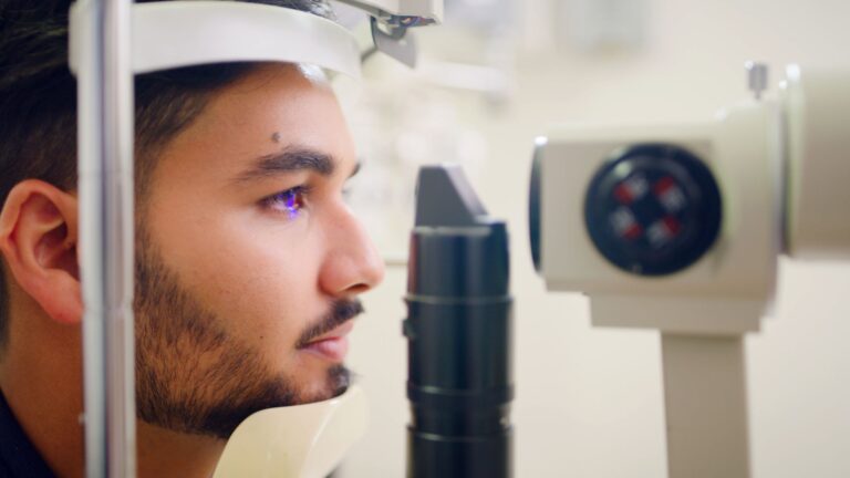
A fundus examination performed by an ophthalmologist allows assessment of the optic disc, which may show signs of damage such as oedema or atrophy, indicative of optic neuropathy.
In ophthalmology, analysis of the optic disc is essential for guiding diagnosis and assessing severity of the pathology.
Magnetic resonance imaging (MRI) is often used to detect associated brain lesions or abnormalities.
Other imaging techniques, such as ocularangiography, may be required to evaluate vascular involvement.
A visual field test is also essential to detect scotomas (areas of vision loss).
Optical coherence tomography (OCT) now allows detailed analysis of the thickness of optic nerve fibres and helps identify degeneration in certain optic neuropathies.
What are the symptoms and visual impact of optic neuropathies?
Symptoms of optic neuropathies may include decreased visual acuity, scotomas (areas of vision loss), colour vision disturbances, and visual discomfort related to inflammation.
People suffering from optic neuropathy may have blurred or reduced vision, sometimes affecting the visual field and daily activities.
This decrease in vision is sometimes described as a persistent haze or blur. In some cases, vision loss may be partial, affecting a specific area of the visual field, or more widespread.
Pain with eye movement may occur, particularly in inflammatory forms such as optic neuritis.
Contrast sensitivity may also be reduced.
The course of the condition may be reversible in some cases (especially inflammatory forms) or may lead to permanent visual damage.
What treatments are available for optic neuropathies?
Optic neuropathies can be managed in different ways, depending on the underlying cause.
In some cases, corticosteroids may be prescribed to reduce inflammation, while in others, surgical intervention may be required, for example in cases of a structural abnormality or a tumour compressing the optic nerve.
The goal of treatment is to limit disease progression and prevent further nerve damage.
Managing cardiovascular risk factors is essential in ischemic forms.
For advanced vision loss, low vision rehabilitation options may help improve quality of life.
Prevention and early detection are crucial, especially for individuals at risk, such as those with a family history of hereditary diseases or conditions like diabetes, which can lead to diabetic retinopathy and optic nerve damage.
Early vision loss may be a warning sign of developing optic neuropathy, and prompt evaluation by an ophthalmologist can help prevent further damage.
Conclusion
Optic neuropathies encompass a range of conditions with multiple causes that can significantly affect vision.
Some forms of optic neuropathies may progress to optic atrophy if not treated promptly.
Early recognition of symptoms, a comprehensive ophthalmological assessment, and appropriate management are essential to limit long-term visual damage.
In the event of sudden or progressive vision loss, prompt specialist consultation is crucial to preserve visual function.
Nous utilisons des cookies pour vous garantir la meilleure expérience sur notre site web. Si vous continuez à utiliser ce site, nous supposerons que vous en êtes satisfait.